Services are available in the following modalities:
Ultrasound, (also referred to as sonography), is an imaging technique that generates high resolution images of internal organs, vessels and tissues in the body using high frequency sound waves that bounce off internal tissues and produce echoes. These echo patterns produce images of the body tissue being examined. Sound waves are sent and received through a small hand held device known as a transducer. The returning sound waves are used to produce the images. Special computer equipment is used to convert these echoes into visual data .Since ultrasound images are captured real time, they can show movement of internal organs and tissues and enable physicians to see blood flow.
Ultrasound based diagnostic imaging techniques are used to visualize muscles and internal organs (including the heart, blood vessels, uterus & bladder), their size, structures and possible pathologies or lesions as well as blood flow through blood vessels. Obstetric Ultrasound is commonly used during pregnancy to assess fetal well being, determine fetal position, verify diagnosis of multiple gestational sacs (twins etc) and rule out ectopic pregnancy. Ultrasound provides information for your physician about the specific soft tissue structure being examined, or about the blood flow in vessels within the soft tissues. Soft tissue structures include a mass in or around a joint, or in a muscle or within your abdomen or pelvis.
The following parts of the body can be imaged using Ultrasound:
• Abdomen: liver, gall bladder, pancreas, spleen major blood vessels.
• Pelvic: reproductive organs, appendix, bladder.
• Obstetrics: rregnancies 1st trimester.
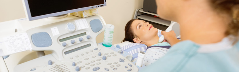
• Superficial structures: thyroid, breast, scrotum.
• Extremities (musculoskeletal): tendons and muscles (shoulder, elbow, knee, ankle, wrist).
Ultrasound uses non-ionizing radiation and is extremely safe. No x-rays are used and there are no known harmful effects or complications.
A completed referral form (requisition) signed by your physician is required to book an appointment for ultrasound. Please bring your requisition and health insurance card with you on the day of your appointment.
Abdomen/Inguinal/Hernia Morning exams |
Avoid fatty dinner. Nothing to eat or drink from midnight the night before (8 hours fasting). Small quantities of clear fluids are permitted. Any medication should be taken as required. |
Abdomen Afternoon exams |
For breakfast you may drink, black tea or coffee (NO MILK OR SUGAR) up to 9 am. Fasting the rest of the day |
Abdomen & Pelvic - Same Visit |
Avoid fatty dinner. Nothing to eat for 8 hours before the exam. Small quantities of clear fluid are permitted. |
Pelvic/Obstetrical |
Finish drinking 4 cups or 1 L (2bottles) of water one hour prior to the exam. DO NOT EMPTY BLADDER. |
Prostate-Transrectal |
The evening before the examination, take Dulcolax Rectal Suppository (purchased at the drug store). Empty bowels before appointments Finish drinking 4 cups or 1 L (2bottles) of water one hour prior to the exam. DO NOT EMPTY BLADDER. |
You will be asked to change into a patient gown prior to your exam. Ultrasound imaging is painless easy and relatively fast. The part of your body being scanned depends on the exam requested by your physician. You will be asked to lie on an exam table and the technologist (sonographer) will spread a lubricant gel on your skin and then press a hand held transducer firmly against your skin on the areas of the requested exam. The gel helps the transducer,(the device that sends sound waves into your body) to make secure contact with your skin and eliminate any air pockets. The transducer is moved back and forth until the desired images are acquired. You will be asked to hold your breath for brief periods of time so that high quality motion free images can be obtained. You may feel some be slight discomfort from pressure as the sonographer guides the transducer over your abdomen, especially if you are required to have a full bladder. The examination takes an average of 30 minutes and you will be able to immediately resume your pre-examination activities.
The results of the ultrasound examination are determined by both the real time (observed during the examination) and the final static images produced. After the examination, this information is reviewed by a radiologist in order to generate a written report for your referring doctor.
Alpha Diagnostic Imaging
1262 Don Mills Road, Suite 206
(Don Mills Rd. & North of Lawrence Ave. E.)
Tel: (416) 510-9977
Fax: (416) 510-3238
Monday - Friday: 9:00 am – 5:00 pm
-
Wheelchair
accesible
Alpha Diagnostic Imaging
2130 Lawrence Ave. E., Suite 300
(Lawrence Ave. E. & Birchmount Rd.)
Tel: (416) 321-2670
Fax: (416) 321-6591
Monday - Thursday: 9:00 am – 6:30 pm
Friday: 9:00 am – 5:00 pm
Saturday: 9:00 am – 2:00 pm
-
Wheelchair
accesible -
Open
weekends